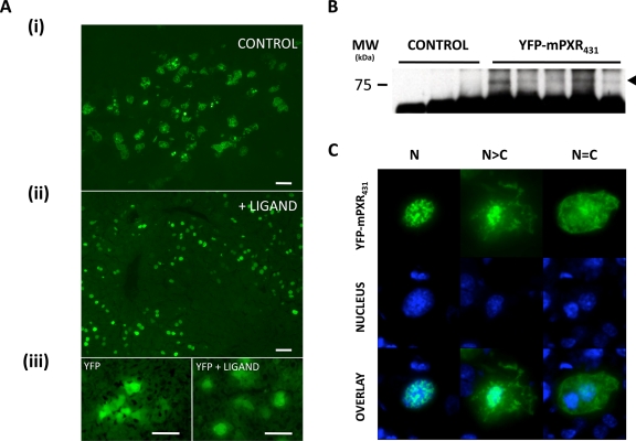
Figure 1. Expression of YFP-mPXR431 in the livers of intact mice.
A. Following the hydrodynamic injection of YFP-mPXR431 expression construct, fluorescence patterns were evident across liver sections of control (i) and ligand (PCN) (ii) treated mice that differed from those of YFP expression alone. B. Immunoprecipitation of YFP-mPXR431 from mouse liver samples indicated a protein band corresponding to the predicted molecular weight of the YFP/PXR fusion protein. C. Subcellular distribution patterns of YFP-mPXR431 expression were categorised into 3 distinct categories. An exclusively nuclear (N), where the fluorescence was confined to the nucleus, a predominantly nuclear (N>C), where a predominant nuclear localisation was evident with some cytoplasmic fluorescence, and an equal distribution (N=C), where nuclear and cytoplasmic fluorescence was evenly distributed across the cell. Nuclei are indicated by blue colour, following Hoechst 33250 staining.

