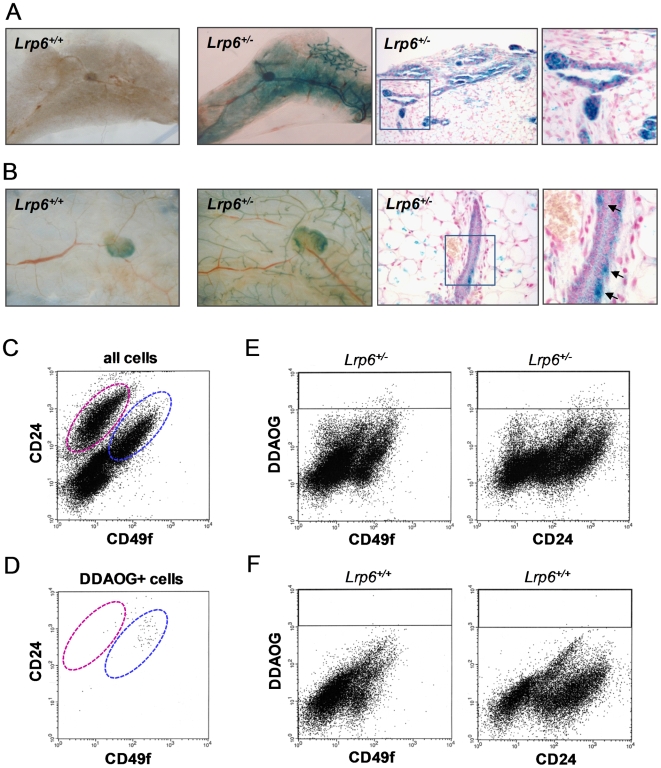
Figure 1. Expression pattern of Lrp6 in the mammary gland.
β-gal expression directed from the Lrp6 promoter in Lrp6+/− mice was used as a surrogate marker for Lrp6 expression. We used the β-gal substrates X-gal and DDAOG to identify cells that express Lrp6. Shown in (A-B) are X-gal-stained mammary whole mounts and 5-µm sections from 2-day-old (A) and 12-week-old (B) female mice. (A) Lrp6 promoter-driven β-gal expression is visible as blue staining in both the basal and luminal mammary epithelium and in the mammary fat pad of newborn mice. (B) In older mice, β-gal-expressing cells are primarily identified within the basal epithelial cell layer and in the mammary fat pad. The arrows indicate typical cells that stained blue with X-gal. No staining was detected in Lrp6+/+ mammary glands, which were used as negative controls for X-gal staining. (C-F) Representative FACS results of DDAOG-stained and CD24/CD49f antibody–labeled mammary epithelial cells. The β-gal-cleaved product of DDAOG has far-red fluorescence and was used to detect cells with Lrp6 promoter–driven β-gal expression. (C) The luminal and basal cell compartments are marked by pink and blue dashed lines, respectively. (D) 80% of the DDAOG-positive cells are found within the basal epithelial cell compartment. (E-F) DDAOG gating strategy. The DDAOG gate is indicated by the black line. The Lrp6+/− sample (E) has 0.33% DDAOG-positive cells; the Lrp6+/+ negative control sample (F) has 0.01% DDAOG-positive cells.

