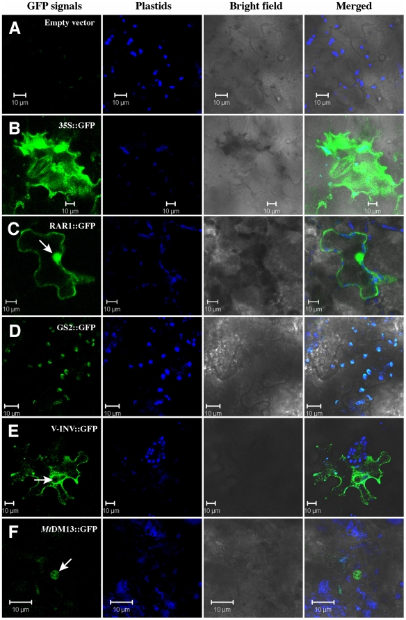
Figure 2. Laser-scanning confocal micrographs showing GFP fluorescence from agroinfiltrated leaf cells.
Katahdin leaves were agroinfiltrated with (A) pK7FWG2 empty vector; (B) 35S::GFP; (C) StRAR1::GFP; (D) StGS2::GFP; (E) StV-INV::GFP; and (F) MtDMI3::GFP. The background fluorescence derived from plastids is in blue color. All the scale bars represent 10 µm. Arrows point to the nucleus in the cells.

