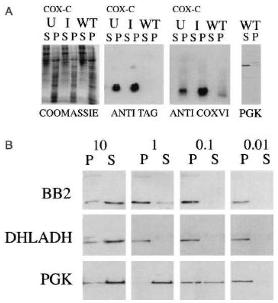
Fig. 4.
A. Cell fractionation of wild type (WT) or COXVI-TY-C (CoxC) expressing trypanosomes in either the presence (I, induced) or the absence (U, uninduced) of tetracycline. The soluble (S) and pellet (P) fractions from cells subjected to freeze–thaw lysis and high-speed centrifugation were probed with either the BB2 antibody or the anti-COXVI antibody. In each case, COXVI localizes to the pellet. A control cyotosolic protein, PGK (Osinga et al., 1985) is detected in the soluble fraction.
B. Crude pellet fractions derived from COXVI-TY-C cells induced with tetracycline were subjected to solubilization in digitonin at concentrations ranging from 0.01 to 10 mg of non-ionic detergent mg−1 trypanosome protein. Soluble (S) protein was separated from pelleted (P) material by centrifugation and blotted to nitrocellulose before being probed with antibodies to the TY epitope, DHLADH or the cytosolic protein PGK.

