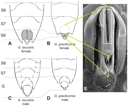
Figure 1. Genital segments in a typical Gerris and in G. gracilicornis.
Schematic drawings of ventral views of abdominal tips of (A) female G. lacustris, (B) female G. gracilicornis, (C) male G. lacustris and (D) male G. gracilicornis. Drawing (B) corresponds to (E) SEM image of the posterior view of a partially inflated female genital segment with gonocoxae 1 spread apart and the ovipositor tube visible. G - genitalia; Gx1 - gonocoxa 1; S6 - sixth segment; S7 - seventh segment; Ov - ovipositor; Vo - vulvar opening. Scale bar: 0.1 mm. (A)-(D) were modified from Andersen [13]. The broken lines schematically indicate the difference between a typical Gerridae and G. gracilicornis in the portion of S8 that is hidden within S7.

