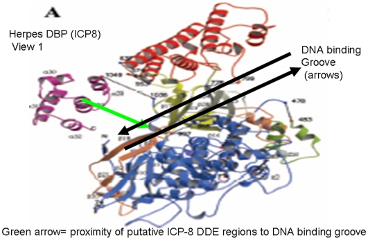
Figure 9. Putative magnesium ion binding regions of the DBP can be localized adjacent to the DNA binding groove of the ICP-8 protein structure.
The partial crystal structure of herpes simplex DBP ICP-8 is shown with experimentally determined DNA binding groove shown, while experimentally determined structures of RAG proteins and other herpes DBP are not solved currently. A black double arrow illustrates the experimentally determined DNA binding groove of ICP-8, while a green arrow indicates the hypothetical position of a bound magnesium ion in ICP-8 as localized by conserved blocks of D and E residues shared with RAG-1 in regions of ICP-8 (Figure 8). This alignment shows that the predicted Mg binding site geometry of ICP-8 is in proximity to the bound DNA as in other structurally characterized DDE enzymes such as RISC. These structural similarities are consistent with and support descent of DBP and RISC proteins from a common precursor DDE recombinase.

