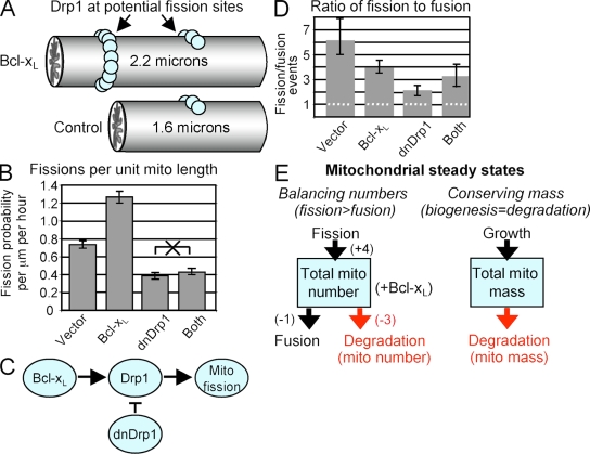
Figure 6.
Bcl-xL induces Drp1-dependent mitochondrial fission. (A) Diagrammatic tubular mitochondrial segments of indicated lengths corresponding to control and Bcl-xL–expressing neurons from Fig. 2 B. These illustrate the distribution of Drp1-containing fission complexes as a function of organelle length. Drp1 molecules (balls) in complexes distributed along mitochondria mark potential sites of future fission events, which implies that the probability of a fission event is proportional to the length of the organelle, with all other conditions being constant. (B) The probability of mitochondrial fission (pfi) per micrometer of mitochondrial length per hour was calculated as described in “Computational methods” (see Table S2, available at http://www.jcb.org/cgi/content/full/jcb.200809060/DC1). P = 0.0002 for all six paired comparisons except for dnDrp1 versus dnDrp1+ Bcl-xL, which was not significant (marked with an X). (C) Diagram illustrating the findings from B. (D) Mitochondrial fission/fusion ratios were calculated from the results shown in Fig. 5 and converted to events per hour. The dotted line marks the theoretical fission/fusion ratio of 1; error bars indicate 25% and 75% bootstrapped quantiles. (E) During steady-state, the number and mass of mitochondria per cell (boxes) must remain equal. This is achieved by an unknown relationship between mitochondrial fission, fusion, biogenesis, and degradation. Parenthetic values for mitochondrial numbers are derived from fission/fusion ratios in Bcl-xL–expressing neurons determined in D.

