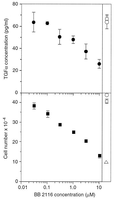
Figure 2.
Inhibition of cell proliferation is correlated with inhibition of TGFα release. (Upper) HMEC strain 184A1 were grown to near confluency and changed to medium containing the indicated concentration of BB-2116 and 20 μg/ml of mAb 225 to prevent ligand uptake by the cells. After 18 hr, the medium was collected and evaluated for TGFα concentration, normalized to 106 cells and ± SD. As a control, cells were also incubated with 50 μM matrix metalloproteinase 3 inhibitor (□) or with no inhibitor (○). (Lower) Cells were split 1:10 into 12-well dishes, and 18 hr later were changed to medium containing the indicated concentration of BB-2116. The medium was changed every 2 days and cells were counted on day 6. Shown are the results of duplicate wells ± SD. Controls are same as Upper as well as 20 μg/ml mAb 225 (▵).

