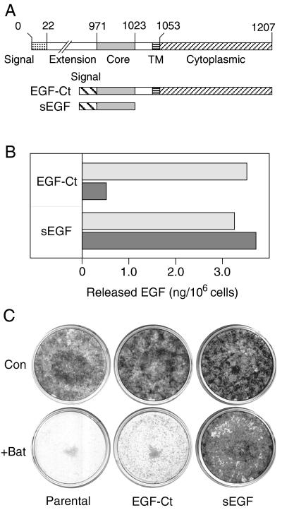
Figure 3.
Batimastat inhibits both release and mitogenic activity of a membrane-anchored, but not a soluble form of EGF. (A) Map of the artificial EGF genes expressed in HMEC. (Upper) The native EGF gene from which the two artificial genes were derived. (B) Cells expressing either the EGF-Ct or sEGF constructs were incubated with 67 nM mAb 225 (to prevent ligand uptake) for 18 hr either without (empty bar) or with (filled bar) 5 μM batimastat. The results are the average of two independent experiments. (C) Parental cells and those expressing the indicated construct were plated at a 1:400 dilution and grown for 2 weeks either with or without 10 μM batimastat (Bat). The medium was changed every 2 days. Cells then were stained with Giemsa.

