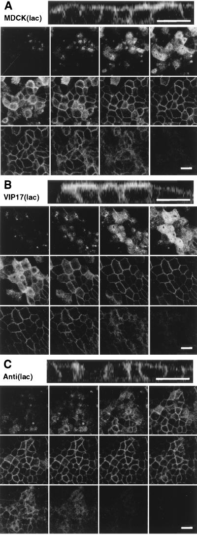
Figure 4.
Localization of a fluorescent GPI -anchored protein in MDCK (lac) cells. Filter-grown MDCK(lac) cells (control cell line, VIP17(lac) clone 48 and Anti(lac) clone A5A3) were induced with 5 mM IPTG for 3 days and then infected with recombinant adenovirus for 1 hr to express YFP-GL-GPI and incubated overnight. Cells were fixed with 4% paraformaldehyde and observed under a Zeiss LSM510 microscope. Twelve optical sections (0.5 μm) along the z axis from the apical cell surface to the base of the cell layer are shown, and the Top image is the x–z view of the same cells. (A) MDCK(lac) cells infected with recombinant adenoviruses to express YFP-GL-GPI. YFP-GL-GPI localized to the apical and basolateral plasma membrane. (B) VIP17(lac) clone 48, induced for 3 days to express epitope-tagged VIP17, was infected with recombinant adenovirus to express YFP-GL-GPI. YFP-GL-GPI shows stronger signal on the apical membrane when compared with control MDCK(lac) cells. (C) MDCK(lac) cells [Anti(lac) clone A5A3], induced to express antisense RNA against VIP17 for 3 days, were infected with recombinant adenovirus to express YFP-GL-GPI. YFP-GL-GPI was mistargeted to the basolateral membrane. (Bar = 20 μm.)

