Figure 5.
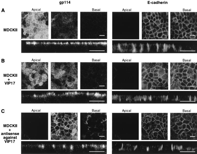
Localization of apical gp114 and basolateral E-cadherin after adenoviral expression of VIP17 or antisense RNA against VIP17 in MDCK II cells. MDCK II cells were grown on filters and infected with recombinant adenoviruses to express VIP17 or antisense RNA against VIP17 for 1 hr and incubated for 3 days. Cells were fixed and processed by using antibodies against gp114 (Left) and E-cadherin (Right) for immunofluorescence microscopy as in Materials and Methods, and then observed under a Zeiss LSM510 microscope. In each set, three optical x–y sections from the apical cell surface to the basal (Upper) and the x–z sections (Lower) are shown. (A) In control MDCK II cells, gp114 was localized to the apical and E-cadherin to basolateral plasma membrane. (B) In cells overexpressing VIP17, the apical surface was enlarged, but the targeting of gp114 and E-cadherin was not changed. (C) In cells expressing antisense RNA against VIP17, gp114 was targeted to the basolateral membrane, whereas the localization of basolateral E-cadherin was unaffected. In some cells of the layer, the lateral cell membranes had changed their appearance. (Bar = 20 μm.)

