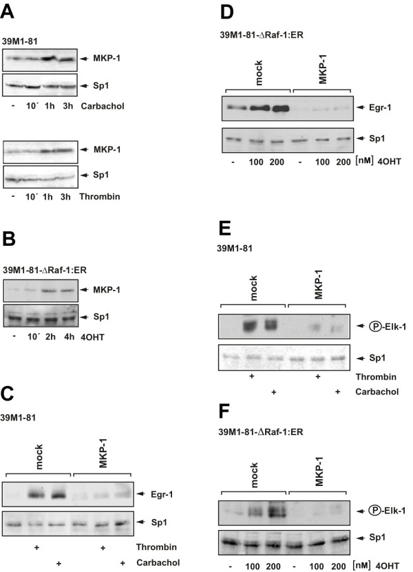
Figure 11.
MKP-1 overexpression prevents Egr-1 expression and Elk-1 phosphorylation in thrombin and carbachol-treated 39M1-81 cells. (A) Western Blot analysis of 39M1-81 cells treated with either thrombin (1 U/ml) or carbachol (100 μM). Nuclear extracts were prepared and subjected to Western blot analysis using an antibody directed against MKP-1. As a control expression of Sp1 is shown. (B) 39M1-81-ΔRaf-1:ER cells were stimulated with 4OHT (100 nM). Western blot analysis of nuclear proteins was performed with an antibody directed against MKP-1. (C) Mock-infected cells or cells infected with a MKP-1 encoding lentivirus were serum-starved for twenty-four hours and then treated with either thrombin (1 U/ml) or carbachol (100 μM) for 1 hour. Nuclear extracts were prepared subjected to Western blot analysis using an antibody directed against Egr-1. (D) MKP-1 controls expression of Egr-1 in 4OHT-stimulated 39M1-81-ΔRaf-1:ER cells. The cells were either mock-infected or infected with a recombinant lentivirus encoding MKP-1. The cells were serum-starved for twenty-four hours and then treated with 4OHT (100 nM) for 2 hours. Nuclear extracts were prepared subjected to Western blot analysis using an antibody directed against Egr-1. (E, F) Mock-infected or cells infected with a lentivirus encoding MKP-1 were serum-starved for twenty-four hours and then treated either with thrombin (1 U/ml) or carbachol (100 μM) for 15 min (E) or with 4OHT (100 nM) for 30 min (F). Nuclear extracts were prepared and subjected to Western blot analysis using an antibody directed the phosphorylated form of Elk-1.

