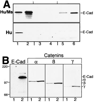
Figure 1.
E-cadherin and catenin expression. (A) Whole-cell lysates (37.5 μg total protein) were probed by Western blotting for E-cadherin by using two different mAbs. (Upper) An antibody that recognizes both human and mouse (Hu/Ms) E-cadherin was used. (Lower) An antibody that recognizes only human (Hu) E-cadherin was used. Lanes: 1, OVCAR-3 human ovarian carcinoma cells (positive control for human E-cadherin); 2, scp2 mouse mammary epithelial cells (positive control for mouse E-cadherin); 3, IOSE-29; 4, IOSE-29neo; 5, IOSE-29preEC; 6, IOSE-29EC. (B) Parental IOSE-29 (lane 1) and IOSE-29EC (lane 2) cell lysates were probed by Western blotting for the 120-kDa E-cadherin protein (100 μg total protein) and the 102-kDa α-catenin, 94-kDa β-catenin, and 86-kDa γ-catenin proteins (20 μg total protein). See Materials and Methods for details of cell lines and antibodies.

