Figure 4.
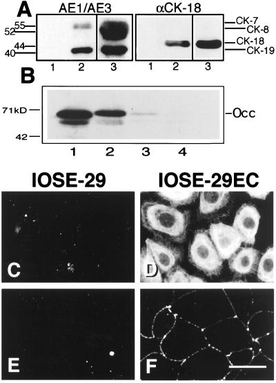
Cytokeratin and occludin expression and localization. (A) Cytoskeletal preparations were probed by Western blotting using a combination of AE1/AE3 antibodies that recognize all cytokeratins except CK-18, or with a specific anti-CK 18 antibody. Lanes: 1, IOSE-29; 2, IOSE-29EC; 3, C4II cervical carcinoma cells (positive control). (B) Whole-cell lysates were probed for the 65-kDa tight-junction protein occludin. Lanes: 1, MDCK kidney epithelial cells (positive control); 2, IOSE-29EC; 3, IOSE-29; 4, 3T3 fibroblasts (negative control). Subconfluent monolayer cultures of IOSE 29 (C and E) and IOSE-29EC (D and F) cells were stained by immunofluorescence for all cytokeratins by using a pan keratin antibody (C and D) and for occludin by using a mAb (E and F). (Bar = 20 μm.)

