Abstract
Tumoral calcinosis is an uncommon lesion, composed of ectopic calcified tissue, most commonly seen in the large joints of the hips, shoulders, and elbows, but may involve the hand and wrist. Patients will often present with localized swelling and reduced mobility around the involved joints. Pain is inconsistent when presenting in the hands or wrists, but the lesions may interfere with daily activities. Multiple variations of the process have been described, ranging from those with no definable etiology (primary), to those associated with disorders (secondary) such as renal insufficiency, hyperparathyroidism, or hypervitaminosis D. The original description of tumoral calcinosis, however, is the familial or hereditary type. Treatment of this process involves optimizing the underlying physiology and complete surgical excision for symptomatic cases.
Keywords: Dialysis, Renal insufficiency, Soft-tissue calcifications, Subcutaneous mass of hand and wrist, Tumoral calcinosis
Introduction
Tumoral calcinosis is a periarticular calcific lesion, which may be encountered by the hand surgeon. The true incidence is unknown, with most descriptions coming from case reports or small series. It can be characterized as a rare condition involving soft-tissue deposits of calcified tissues, most commonly around larger joints, occasionally involving the joints of the hand or wrist. Patients may be referred to the hand surgeon with solitary or multiple masses in the hand or wrist to establish a diagnosis as well as render definitive treatment. This report presents an example of such a case and discusses the pathogenesis and treatment as it relates to the hand surgeon.
Case Report
A 51-year-old right-hand-dominant Caucasian female was seen following a 15-month history of multiple, slowly enlarging masses on her right hand and wrist. The lesions caused mild discomfort and occasionally interfered with activities. Radiographs were obtained and revealed multiple calcifications of unclear etiology, which prompted the hand surgery referral.
Her medical history was significant for renal insufficiency and a failed renal transplant, requiring hemodialysis, right breast cancer, hypertension, and hyperlipidema. Her breast cancer treatment was completed 6 months prior to her presentation and did not require chemotherapy or radiation. Her medications were consistent with her medical conditions.
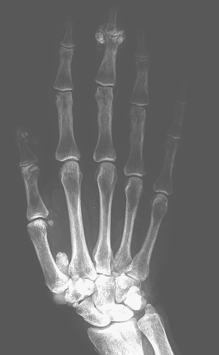
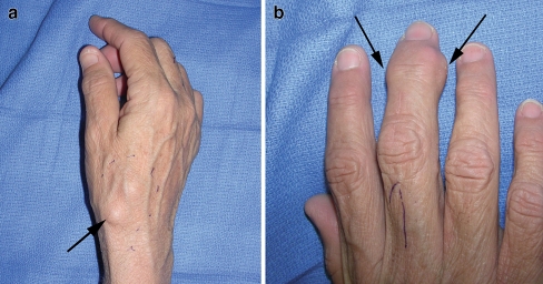
Her hand exam was significant for multiple mildly tender masses, the most prominent of which was located along the volar aspect of the middle finger near the distal interphalangeal joint. Additional masses were noted along the dorsal radial aspect of the base of the thumb metacarpal and the volar aspect of the index and small fingers in the region of the metacarpophalangeal joints (Fig. 1a and b). Radiographs revealed calcified masses in these regions (Fig. 2).
Figure 1.
a Preoperative appearance of a lesion at the dorsal radial aspect of the base of the thumb. b Preoperative appearance of a lesion along the volar aspect of the middle finger.
Figure 2.

Radiographic appearance of the right hand.
It was felt these most likely represented a benign processes related to her renal disease. The patient desired a definitive diagnosis secondary to her history of breast cancer, so excision of the two most symptomatic lesions were completed with regional anesthesia.
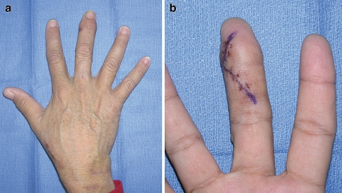
The specimen was not encapsulated, but was easy to remove. It appeared as a yellowish, white chalky substance and when sectioned, contained milky liquid and cystic spaces (Fig. 3a and b). In the final pathology, a diagnosis of soft-tissue masses consistent with tumoral calcinosis was reported. Her healing of these sites was uneventful, and although she developed enlargement of the other existing lesions, she elected not to undergo further treatment (Fig. 4a and b).
Figure 3.
a Intraoperative appearance of the thumb lesion. b Intraoperative appearance of the middle finger lesion. c Gross specimen dissected from the thumb lesion.
Figure 4.
a Postoperative appearance of the hand. b Postoperative appearance of the middle finger.
Discussion
Tumoral calcinosis is an uncommon disorder, but may be encountered in the hand and wrist. The phrase tumoral calcinosis was originally described in 1943 by Inclan [5]. The original condition was described in 1899, when Duret described the process in siblings with multiple calcifications in the hips and elbows [1]. A Medline search of English language literature revealed multiple descriptions of tumoral calcinosis, and multiple clinical entities thought to be the cause of the condition [2–14]. The condition is described as a familial type, seen in young, otherwise healthy individuals in the second or third decade of life and frequently affects multiple siblings. It is also described as a spontaneous entity or secondary to chronic renal insufficiency, hyperparathyroidism, hypervitaminosis D, and other metabolic disorders. It is typically associated with high calcium and phosphate levels.
As originally described, tumoral calcinosis is a heredity condition or familial type. The term is now routinely and erroneously used to describe any soft-tissue periarticular calcification. Histologically, these lesions appear the same [9], which explains why periarticular calcifications are often called tumoral calcinosis, regardless of the etiology. Most of the described cases are not tumoral calcinosis by the original definition, but periarticular calcifications. Fortunately, the treatment is the same for all conditions [10].
When presented with a soft-tissue mass, imaging studies are usually obtained. Plain radiographs typically reveal periarticular calcifications without involvement of the underlying joints. CT scans will often reveal cystic spaces within the calcified masses, especially in larger lesions and ultrasound imaging will reveal fluid accumulation within the cystic space [2]. When underlying osseous changes are noted, the differential diagnosis should include other conditions, including malignant processes.
As with our patient, lesions may appear in other locations. Following diagnosis, medical treatment of the underlying conditions will decrease the development of new lesions and symptomatic lesions can be excised.
References
- 1.Duret MH. Tumeurs et singuliere des bourses (endotheiomes peut etre d'origine paraasitair). Bull Mem Soc Anat Paris 1899;74:275–7.
- 2.Franco M, Van Elslande L, Passeron C, Verdier JF, Barrillon D, Cassuto-Viguier E, et al. Tumoral calcinoisis in hemodialysis patients. A review of three cases. Rev Rheum Engl Ed 1997;64:59–62. [PubMed]
- 3.Hamada J, Tamai K, Ono W, Saotome K. Uremic tumoral calcinosis in hemodialysis patients: clinicopathological findings and identification of calcific deposits. J Rheumatol 2006;33:119–26. [PubMed]
- 4.Huang YT, Chen CY, Yang CM, Yao MS, Chan WP. Tumoral calcinosis-like metastatic calcification in a patient on renal dialysis. J Clin Imaging 2006;30:66–8. doi:10.1016/j.clinimag.2005.06.024. [DOI] [PubMed]
- 5.Inclan A. Tumoral calcinosis. JAMA 1943;121:490–5.
- 6.Jones G, Kingdon E, Sweny P, Davenport A. Tumoral calcinosis and calciphylaxis presenting in a dialysis patient. Nephrol Dial Transplant 2003;18:2668–70. doi:10.1093/ndt/gfg367. [DOI] [PubMed]
- 7.Kim HS, Suh JS, Kim YH, Park SH. Tumoral calcinosis of the hand: three unusual cases with painful swelling of the small joints. Arch Pathol Lab Med 2006;130:548–51. [DOI] [PubMed]
- 8.Murai S, Matsui M, Nakamura A. Tumoral calcinosis in both index fingers: a case report. Scand J Plast Reconstr Surg Hand Surg 2001;35:433–5. doi:10.1080/028443101317149426. [DOI] [PubMed]
- 9.Olsen KM, Chew FS. Tumoral calcinosis: pearls, polemics, and alternatives. Radiographics 2006;26:871–85. doi:10.1148/rg.263055099. [DOI] [PubMed]
- 10.Polykandriotis EP, Beutel FK, Horch RE, Grunert J. A case of familial tumoral calcinosis in a neonate and review of the literature. Arch Orthop Trauma Surg 2004;124:563–7. doi:10.1007/s00402-004-0715-0. [DOI] [PubMed]
- 11.Savaci N, Avunduk MC, Tosun Z, Hosnuter M. Hyperphosphatemic tumoral calcinosis. Plast Reconstr Surg 2000;105:162–5. doi:10.1097/00006534-200001000-00027. [DOI] [PubMed]
- 12.Tadjalli HE, Kessler FB, Abrams J. Tumoral calcinosis of the triangular fibrocartilage complex: a case report. J Hand Surg 1997;22A:350–3. [DOI] [PubMed]
- 13.Tezelman S, Siperstein AE, Duh QY, Clark OH. Tumoral calcinosis. Controversies in the etiology and alternatives in the treatment. Arch Surg 1993;128:737–44. [DOI] [PubMed]
- 14.Tong MKH, Siu YP. Tumoral calcinosis in end stage renal disease. Postgrad Med J 2004;80:601. doi:10.1136/pgmj.2003.017608. [DOI] [PMC free article] [PubMed]


