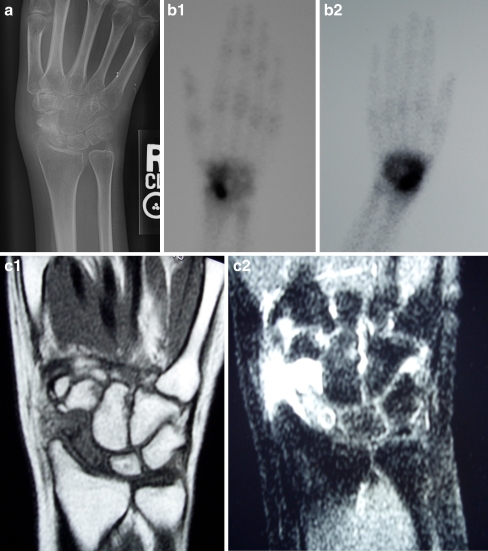
Figure 1.
a Plain radiographs demonstrate a sclerotic lesion in the proximal scaphoid pole. Radiocarpal arthrosis with narrowing of the radiocarpal joint space and overgrowth of the radial styloid is noted. b Bone scan demonstrates intense uptake at the scaphoid with diffuse uptake across the carpus on delayed imaging. c MRI demonstrating homogeneous hypointense signal of the scaphoid on T1 sequences and hyperintense signal on T2 images with focal proximal hypointensity.

