Figure 1.
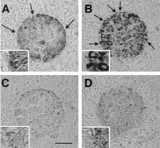
Cytochemical detection of H2O2 production in 3T3 cells. Cells were irradiated with blue light (450–490 nm at 6.3 W/cm2) for 20 min in saline containing AEC followed by thorough washing to remove large nonspecific crystals. (A) In normal saline, irradiated cells (circular area) displayed moderately dark reaction product (polyAEC) with a punctate distribution. (B) In cells pretreated with catalase, staining was darker and clearly localized in the cytoplasm. (C) Staining was markedly attenuated in cells fixed for 1.5 hr in buffered 4% paraformaldehyde solution before irradiation. (D) Staining was reduced and punctate in cells pretreated with vitamin C. Arrows denote cells with partial staining. [Bar = 100 μm and 50 μm (Inset).]

