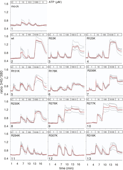
Fig. 2.
Potency of ATP to induce calcium flux in HEK cells transiently transfected with P2X7 variants. HEK cells were co-transfected with expression constructs for mRFP and wild-type or mutant P2X7 purinoceptors. Twenty hours post-transfection, cells were loaded with the calcium-sensitive fluorochrome Fura-2 before live cell imaging by fluorescence microscopy. Images were captured every 5 s. At the indicated times, the perfusion buffer (37°C) was changed to subject cells to increasing doses of ATP. Ratio images (340/380 nm) were constructed pixel-by-pixel and single cell tracings were captured using the Openlab software. Gray lines show single cell tracings, red lines the calculated mean

