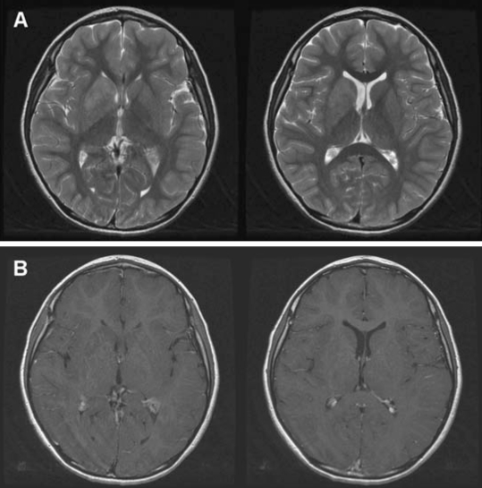
Figure 2.
Magnetic resonance imaging showing an ill-defined mass lesion involving the right basal ganglia. The lesion was located in the putamen, globus pallidus, head of caudate, and the anterior limb of the internal capsule. (A) Axial T2- weighted images, and (B) axial T1-weighted contrast enhancement images.

