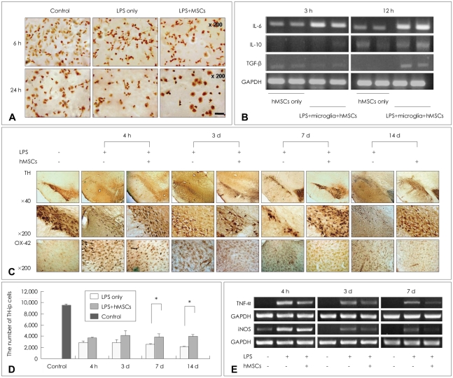
Fig. 2.
Coculturing hMSCs with LPS-stimulated microglia in a Transwell culture chamber system decreased microglial activation and increased the expressions of IL-6, IL-10, and TGF-β. To identify soluble factors associated with modulation of microglial activation, we analyzed the expressions of IL-6, IL-10, and TGF-β in hMSCs cocultured with LPS-stimulated microglia and in hMSCs alone. The inclusion of hMSCs significantly decreased the number of process-bearing activated microglia at 6 and 24 h following hMSC treatment (A). When hMSCs were cocultured with LPS-stimulated microglia, IL-6 expression was significantly increased at 3 and 12 h, and the expressions of IL-10 and TGF-β at 12 h were significantly higher than those with hMSCs alone (B). Immunohistological evaluation of protective effect of hMSCs against LPS-induced damage to dopaminergic neurons in the SN. hMSC treatment considerably reduced the loss of TH-ir cells and microglial activation induced by LPS stimulation in the SN (C, Scale bar: 100 mm). Stereological analysis revealed that hMSC treatment significantly decreased the loss of TH-ir cells at 7 and 14 days following LPS stimulation (D, *p<0.05). The administration of hMSCs significantly down-regulated the LPS-induced increase in the expressions of TNF-α and iNOS mRNA at 3 days after LPS stimulation (E). hMSCs: human mesenchymal stem cells, LPS: lipopolysaccharide, IL: interleukin, TGF-β: transforming growth factor β, SN: substantia nigra, TNF-α: tumor necrosis factor-α.

