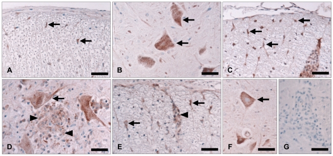
Fig. 2.
Immunohistochemical staining of Epo in the spinal cords of normal control rats (A and B) and rats at the peak (day 12 PI)(C and D) and recovery (day 21 PI)(E and F) stages of EAE. Epo was weakly detected in some glial cells (A, arrows) and neurons (B, arrows) in the spinal cords of normal control rats. Glial cells at the peak stage of EAE showed increased immunoreactivity for Epo (C, arrows). Epo-positive inflammatory cells (D, arrowheads) and neurons (D, arrow) were detected in the parenchyma. At the recovery stage of EAE, some inflammatory cells (E, arrowhead), glial cells (E, arrows) and neurons (F, arrows) were positive for Epo. Tissue from a rat at the peak stage of EAE stained without the primary antisera showed no staining (G). Counterstaining was with hematoxylin. Scale bars=40 µm. Epo: Erythropoietin, PI: post-immunization, EAE: experimental autoimmune encephalomyelitis.

