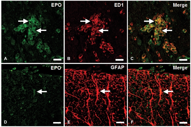
Fig. 3.
Immunofluorescent co-localization of Epo (A and D) with ED1 (B) and anti-GFAP (E) in the spinal cords of rats at the peak stage of EAE (day 12 PI). A large amount of Epo (A, green; arrows) was immunostained in ED1-positive macrophages (B, red; arrows)(C, merge; arrows). Epo immunoreactivity (D, green; arrow) was co-localized in GFAP (E, red; arrow)-positive astrocytes (F, merge; arrow). Scale bars: A-F, 20 µm. Epo: Erythropoietin, anti-GFAP: anti-glial fibrillary acidic protein, EAE: experimental autoimmune encephalomyelitis, PI: post-immunization.

