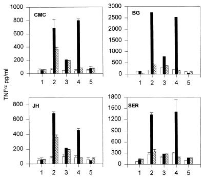
Figure 1.
Enhancement of TNFα release from coincubation of stimulated THP-1 cells and human neutrophils. Representative data are shown from two separate experiments by using two neutrophil donors per day. Initials in top left hand corner of graphs refer to individual donors. One × 106 THP-1 cells (hatched bars) and 4 × 10⁶ neutrophils isolated from individual donors were added to tissue culture wells either separately (open bars) or as a mixture of both populations (closed bars). Numbers along the horizontal axes indicate different conditions. Cells were incubated for 4 h at 37°C either with no further additives (position 1), with 1 μg/ml LPS plus 10-5 M FNLP (position 2), 10 μM CE-2072 (position 3), 10 μM GI-1 (position 4), or 10 μM CE-2072 plus 10 μM GI-1 (position 5). Supernatants were recovered and assayed for TNFα by ELISA. For each donor data represent mean TNFα levels ± SD from two tissue culture wells with each well run in duplicate in ELISA (n = four observations per donor).

