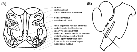
Figure 2.
(A) Schematic of the anatomical structures located in the caudal medulla where the lesion was found in MRI and its sagittal level of the brainstem. The round gray areas indicate the presumed locations of the lateral vestibulospinal tract in the lower medulla oblongata, and the area with diagonal lines indicates the lesion in the patient. (B) Straight line indicates the level of the caudal medulla.

