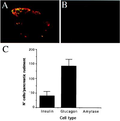
Figure 2.
Immunohistological and quantitative analysis of E11.5 rat pancreatic rudiments. (A) Immunostaining with anti-glucagon (red fluorescence) and anti-insulin (green fluorescence) antibodies. (B) The lack of positive staining for amylase staining indicates the absence of development of acinar cells. (C) Quantification of the number of insulin-, glucagon-, and amylase-expressing cells in the E11.5 rat pancreas. Each data point represents the mean ± SEM of 15 pancreatic rudiments.

