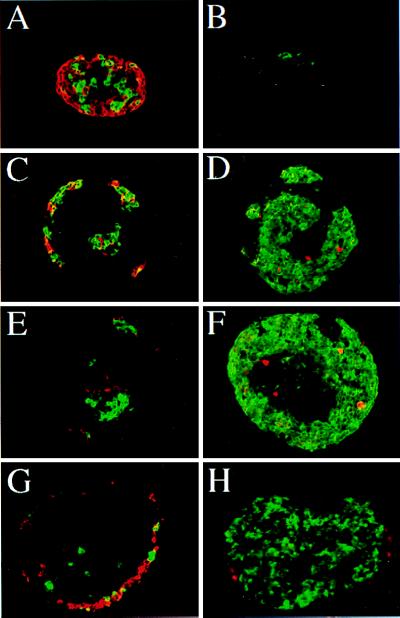
Figure 4.
Immunohistological analysis of pancreatic epithelium grown in vitro in the presence of FGFs. E11.5 mesenchyme-free pancreatic epithelia were grown in collagen gels for 7 days in the absence (A and B) or presence of 50 ng/ml of either FGF-1 (C and D), FGF-7 (E and F), or FGF-10 (G and H). The development of the endocrine tissue (A, C, E, and G) was evaluated after anti-glucagon (red fluorescence) and anti-insulin (green fluorescence) staining. The development of the exocrine tissue (B, D, F, and H) was evaluated after anti-amylase (green fluorescence) staining. The effect of FGF-1 and FGF-7 on acinar cell proliferation (B, D, and F) was analyzed by staining with the antimitotic proteins, mpm2 antibody (red fluorescence). Magnifications: A and B, ×600; C-H, ×400.

