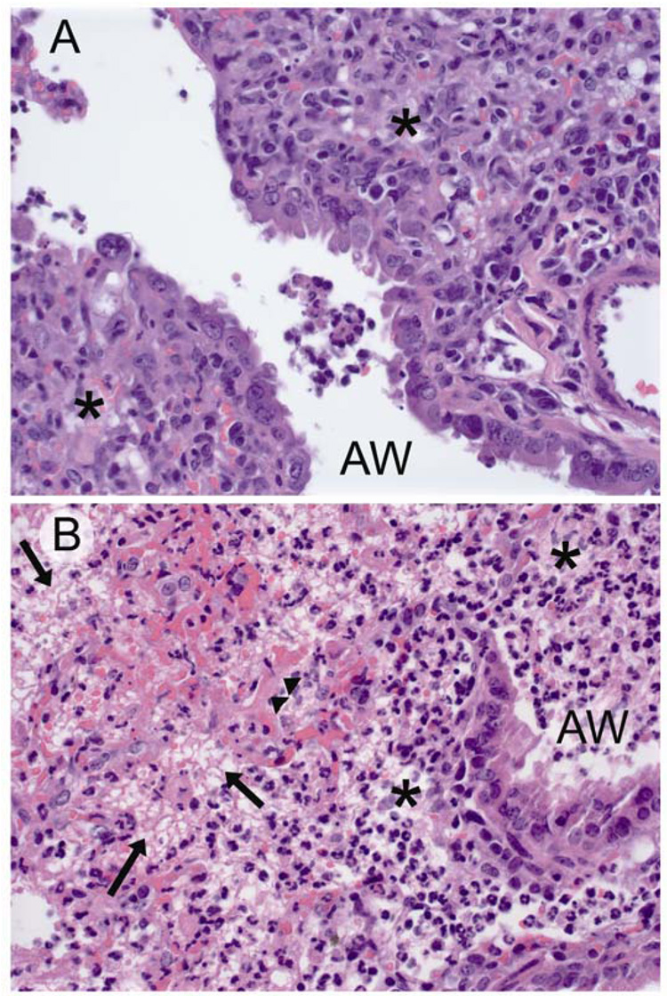
Figure 6.
Histopathology of mice treated for secondary bacterial pneumonia. Representative hematoxylin and eosin stained sections (40X) from of terminal airways and interstitium of lungs of mice infected with influenza virus PR8, then challenged 7 days later with S. pneumoniae and treated with either A) clindamycin or B) ampicillin. Lungs were taken 24 hours after start of treatment. in A) mononuclear inflammation (*) can be seen in the interstitium with a few admixed neutrophils. In B) fibrinonecrosis of the insterstitium is evident with effacement of the alveoli by lacy eosinophilic fibrin strands (arrows), copious infiltrates of both viable and degenerate neutrophils (*), and necrotic alveolar walls (arrowheads). AW = airway.

