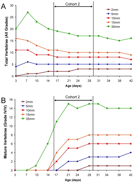
Figure 3.
Timing of postnatal tail vertebral development by using microcomputed tomography. A 35-mm segment of tail was scanned by using microCT and vertebrae (n = 18 mice for each age group [strain factor collapsed]) in measured segments of tail (2, 5, 10, or 15 mm) determined. (A) The total number of vertebrae in the 35-mm sections decreased over time, after a peak within the first week. Counts in the 10- to 15-mm segments decreased with increasing age. Counts in the shortest sections (2 and 5 mm) increased for the first 2 to 3 wk of age and then plateaued, with consistent counts through day 42. (B) The number of vertebrae that had end plates (MV; grades IV and V) increased as a function of age for each measured segment. On average, MV were not noted in the most distal 2 mm before day 21; however, MV are present in all sections greater than or equal to 5 mm before day 21.

