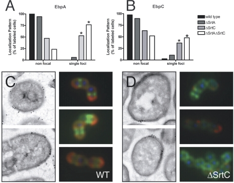
FIG. 5.
Pilus subunits accumulate focally in the absence of SrtC. (A and B) Quantification of EbpA (A) or EbpC (B) immunofluorescent labeling of whole E. faecalis OG1X wild-type or sortase mutant cells grown to stationary phase. *, P < 0.0000001 by Fisher's exact test. (C and D) EbpA labeling of wild-type (WT) (C) or ΔSrtC bacteria (D) and localization by electron microscopy (left panels). Representative images of whole-cell immunofluorescence labeling of EbpA (red), DNA (blue), and cell wall (green) are also shown (right panels).

