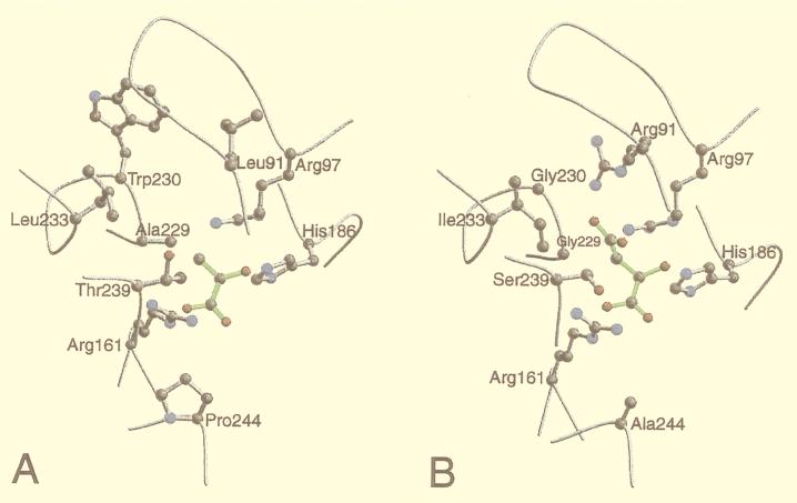
Figure 3.
Plots of the active site environments in the 3D models of T. vaginalis lactate (A) and malate (B) dehydrogenases. The active site environment includes all of the residues that have at least one atom within 5 Å of at least one substrate atom. The backbone is traced in gray. The conserved Arg161 and His186 residues, presumed to be crucial in catalysis, and the residues that differ in type between the two sequences are shown in the ball-and-stick representation. The substrate molecules are shown in green. The plots were drawn with the programs molscript (41) and raster3d (42).

