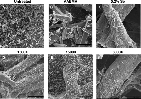
FIG. 4.
SEM analysis of P. aeruginosa (A to C) and S. aureus (D to F) biofilm formation on untreated, AAEMA-coated, or Se-MAP-coated (0.2% selenium) cellulose discs. Biofilms were allowed to form as described for Fig. 3. After 24 h of incubation at 37°C, the discs were fixed, dried, affixed to aluminum mounts, and sputter coated with platinum and palladium. Observations were performed at 6 to 7 kV with a scanning electron microscope. Five fields of view were examined from randomly chosen areas from the optical surface of each sample at magnification of ×1,500 for untreated and AAEMA-coated discs and at ×5,000 for the 0.2% selenium Se-MAP-coated discs. Each experiment was conducted in triplicate. Representative fields of view are shown. Bars, 20 μm. Crystals visible in panels B and F are artifacts of fixation.

