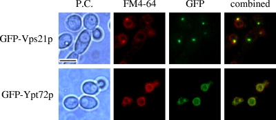
FIG. 4.
Vps21p and Ypt72p localize to endosomal and vacuolar compartments, respectively. Cells expressing the GFP-VPS21 or GFP-YPT72 fusion were pulse-labeled with FM4-64 and chased in fresh medium for 20 min (top) to label endosomes or for 60 min (bottom) to label vacuoles. Cells were then observed by phase-contrast (P.C.) microscopy (left column) and epifluorescence microscopy with a TRITC filter for FM4-64 (second column from the left) or a fluorescein isothiocyanate filter for GFP (third column). The combined FM4-64-GFP images are also shown (fourth column). FM4-64 colocalized with GFP-Vps21p and GFP-Ypt72p in all cells with detectable levels of these fusion proteins (>95% of the cells had detectable GFP-Vps21p expression; >90% of the cells had detectable levels of GFP-Ypt72p). Bar = 5 μm.

