
FIG. 1.
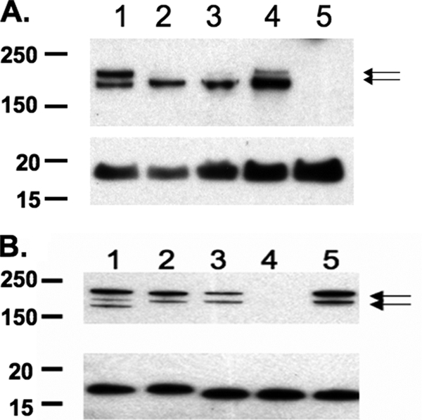
Western blots using a rabbit anti-HrpA antibody and monoclonal antibody 2C3 that binds to the outer membrane protein H.8. H.8 was used as the loading control for the individual samples (A and B, lower images). (A) Lysates of strain NMB siaA-D grown under aerobic conditions (lane 1) or anaerobic conditions for 1 (lane 2), 2 (lane 3), and 24 h (lane 4); lane 5 shows the growth of NMB siaA-D hrpA. The normalized 180-kDa band in lane 4 was significantly upregulated compared to the normalized 180-kDa band in lane 1. (B) Lysates of strains MC58 siaD (lane 1), C311 (lane 2), NMB siaA-D (lane 3), NMB siaA-D hrpA, (lane 4), and a lysate of NMB siaA-D hrpA complemented with hrpA (lane 5) grown aerobically. Arrows indicate the HrpA-specific (220- and 180-kDa) bands.