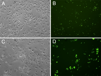
FIG. 1.
Analysis of Y. pestis replication in activated macrophages by microscopy. BMDMs infected with KIM5+/GFP were activated with IFN-γ and incubated for 6 h (A and B) or 24 h (C and D) before microscopic examination. One hour before examination, IPTG was added to induce de novo expression of GFP in viable intracellular bacteria. (A and C) Representative phase-contrast microscopy images. (B and D) Representative fluorescence microscopy images.

