FIG. 3.
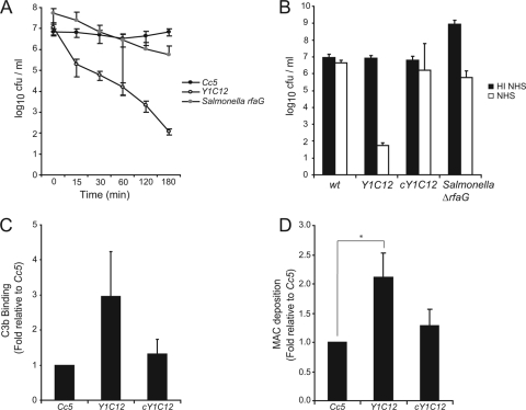
Characterization of the serum-sensitive Tn mutant of Cc5. (A) Total CFU present after incubation of Cc5 (black circles), Y1C12 (white circles), or an S. enterica serovar Typhimurium rfaG mutant (gray circles) in 10% NHS for 0, 15, 30, 60, 120, and 180 min at 37°C. (B) Total CFU present after incubation of Cc5, Y1C12, cY1C12, or an S. enterica serovar Typhimurium rfaG mutant in 10% HI NHS (black) or 10% NHS (white) for 180 min at 37°C. (C) Binding of C3b by Cc5, Y1C12, or cY1C12 after 30 min incubation with C7-depleted NHS. FACS analysis was performed using anti-C3c polyclonal antiserum to quantify the level of C3b on the bacterial surface. (D) MAC deposition on Cc5, Y1C12, or cY1C12 after 15 min of incubation with NHS (longer incubation leads to significant lysis). FACS analysis was performed using mouse anti-C5B-9 primary and anti-mouse-FITC secondary Abs to quantify the level of MAC insertion into the bacterial surface. The statistical significance of the difference in MAC insertion between Y1C12 and wt C. canimorsus is given, with * indicating a P value of <0.05 using a two-tailed unpaired Student's t test. Results are the means of at least three independent experiments ± standard deviations (bars). cY1C12, Y1C12 mutant bacteria complemented with the gtf gene in trans.

