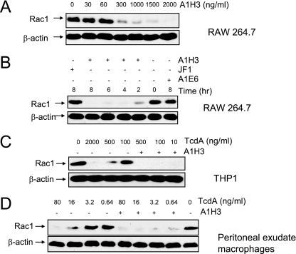
FIG. 2.
A1H3-dependent enhancement of the glucosyltransferase activity of TcdA. RAW 264.7 or THP1 cells were treated with TcdA in the presence or absence MAbs. Protein lysates were separated by sodium dodecyl sulfate-polyacrylamide gel electrophoresis, transferred to a nitrocellulose membrane, and probed with anti-beta-actin and anti-Rac1 (MAb 102) antibodies. (A) RAW 264.7 cells were treated with TcdA (0.4 ng/ml) in the presence of the indicated doses of A1H3 for 4 h. (B) RAW 264.7 cells were incubated with TcdA (0.4 ng/ml) with or without the indicated MAbs for the times indicated. (C) THP1 cells were incubated with various doses of TcdA with or without A1H3 for 4 h. (D) Mouse peritoneal exudate macrophages were exposed to the indicated amounts of TcdA with or without A1H3 for 5 h.

