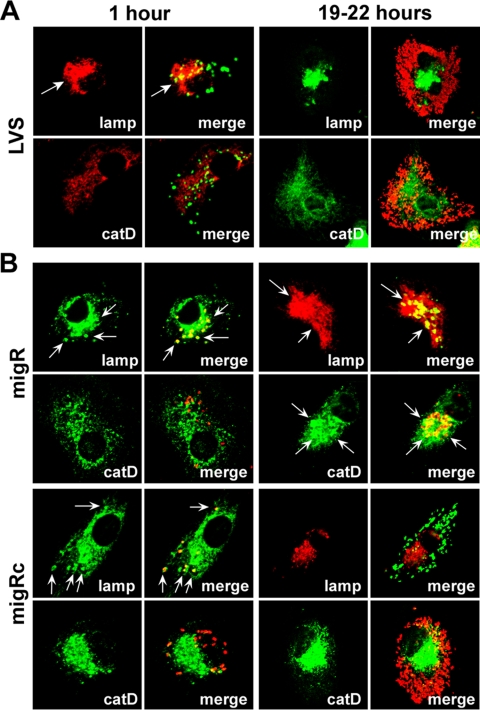
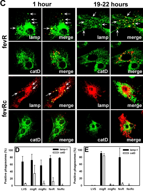
FIG. 6.
Composition of migR and fevR mutant phagosomes in MDMs. (A to C) Representative confocal sections of MDMs infected for 1 h or 19 to 22 h (overnight) at 37°C with LVS (A), the migR mutant or its trans-complemented migRc strain (B), or the fevR mutant and its trans-complemented fevRc strain (C). In each case, samples were stained to detect bacteria and lamp-1 (lamp) or cathepsin D (catD), as indicated. Arrows indicate positive phagosomes. (D to E) Percentage of bacteria inside MDMs that were infected for 1 h (D) or overnight (E) that were inside lamp-1- or cathepsin D-positive phagosomes. Data are the averages ± the standard errors of the means of the results from three independent experiments performed in triplicate.


