FIG. 6.
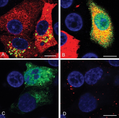
Effects of DN rab7 on LDL trafficking and FMDV infection. (A and B) IBRS-2 cells were transfected to express wt rab7 (A) or DN rab7 (B) and infected with FMDV. Panels A and B show representative cells from one experiment. Cells expressing the rab protein (green) were identified by the EGFP tag. FMDV-infected cells (red) were identified as for Fig. 1. Cells expressing either wt or DN rab7 were scored for infection as for Fig. 3. (Quantification of these data is shown in Fig. 7.) (C) IBRS-2 cells were transfected to express DN rab7. (D) Uptake of Alexa-568-labeled LDL for the same cells as shown in panel C. In the nonexpressing cells, LDL (red) accumulated in larger perinuclear vesicles characteristic of LE, whereas in the cells expressing DN-rab7, LDL labeling was confined to smaller, more peripheral vesicles. The cell nuclei are shown in blue. Bars, 10 μm.

