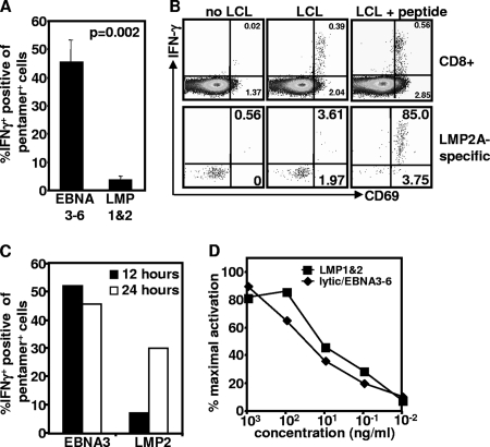
FIG. 3.
Recognition of endogenous antigen by EBV-specific T cells. PBMC were stimulated with autologous LCL at a 20:1 responder-to-stimulator ratio in the presence of brefeldin A. (A) IFN-γ expression was assessed using intracellular cytokine assay. Data represent the means and standard errors of the results for IFN-γ-producing CD8+ T cells from PBMC of six donors in each group following incubation with autologous LCL for 12 h. (B) CD69 and IFN-γ expression by total CD8+ T cells or HLA pentamer-specific CD8+ T cells was assessed using intracellular cytokine assay. Representative data are from CD8+ T cells or LMP-2a-specific T cells from a single donor following stimulation with and without LCL, pulsed with and without an LMP-2a-specific epitope, CLGGLTMV. Data in the upper right quadrant are the percentages of CD69+ IFNY+ cells. Data in the lower right quadrant are the percentages of cells producing CD69 alone. (C) Representative data from single donors showing recognition of LCL by EBNA3-specific T cells recognizing the epitope FLRGRAYGL or LMP2a-specific T cells recognizing the epitope CLGGLLTMV after 12 or 24 h in the presence of brefeldin A for the previous 6 h. (D) EBV-specific T-cell avidity. PBMC from EBV-seropositive donors were stimulated overnight with 10-fold serial dilutions of HLA-matched EBV lytic antigen, EBNA3-6, EBNA1, or LMP-1/2 peptides. Activation of CD8+ T cells was assessed by the upregulation of the early activation markers CD137 and CD69.

