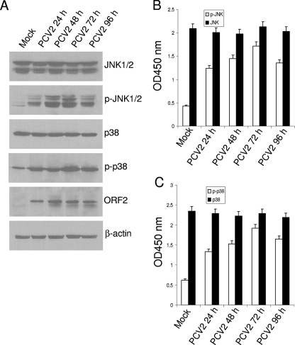
FIG. 1.
PCV2 infection activates JNK1/2 and p38 MAPK signaling pathways in the cultured cells. (A) Whole-cell lysates from PK15 cells after infection with PCV2 strain BJW at an MOI of 1 TCID50. PCV2-infected cells were harvested at 24, 48, 72, and 96 h postinfection, and whole-cell lysates were prepared and resolved by SDS-PAGE, transferred to nitrocellulose membranes, and immunoblotted. The protein levels of JNK1/2 and p38 and their phosphorylated forms, as well as PCV2 viral capsid protein, were analyzed. The amounts of β-actin were also assessed to monitor the equal loadings of protein extracts. (B and C) JNK1/2 (B) and p38 MAPK (C) activation induced by PCV2 infection was determined by using FACE assay. PK15 cells were fixed at the indicated time points with 4% formaldehyde and incubated with antibodies directed against JNK1/2 or p38 or their phosphorylated forms followed by HRP-conjugated immunoglobulin G antibodies. JNK1/2 and p38 and their phosphorylated forms were each assayed in triplicate. Cell numbers were normalized by using crystal violet. These results are representative of three independent experiments. Values are means ± the SD from triplicate wells. p-, Phosphorylated.

