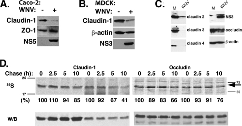
FIG. 4.
WNV causes degradation of claudin family proteins. Control and WNV-infected Caco-2 (A) or MDCK (B) cell lysates were analyzed by Western blotting to determine the levels of claudin-1, ZO-1, and NS5 (Caco-2) or claudin-1, β-actin, and NS3 (MDCK). (C) Control and WNV-infected Caco-2 cell lysates were analyzed by Western blotting to determine the levels of claudin-2, -3, and -4, occludin, NS3, and β-actin. (D) Control and WNV-infected cells were metabolically labeled with [35S]methionine and chased for the indicated times. Claudin-1 was immunoprecipitated from the lysates, and supernatants of claudin-1 immunoprecipitates were used for occludin immunoprecipitation. Protein complexes were resolved by SDS-PAGE and transferred onto PVDF membranes. Claudin-1 and occludin half-lives were quantified by autoradiography. The positions of the molecular weight marker bands are shown for both gels; an arrow points to the occludin band. Western blot analysis of the membranes with respective antibodies confirmed equal amounts of protein immunoprecipitation in all samples.

