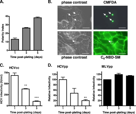
FIG. 1.
Effect of HepG2 polarity on HCV entry. (A) HepG2 cells were grown for 1, 3, or 5 days, fixed in 3% paraformaldehyde, and stained for the BC-expressed marker MRP2. The polarity index was assessed by quantifying the number of MRP2-positive BC per 100 cell nuclei for five fields of view on three replicate coverslips. (B) The BC in polarized HepG2 cells were assessed for both “barrier” and “fence” functions. Cells were incubated with either C6-NBD-SM, to measure fence function, or CMFDA, which measures barrier function. Restriction of C6-NBD-SM to the basal plasma membrane and restriction of CMFDA to the BC indicate that polarized HepG2 cells have functional TJs. (C) HCVcc J6/JFH was used to infect HepG2-CD81 cells at 1, 3, or 5 days postplating. Infected cells were visualized after 72 h (NS5A staining), and infectivity (focus forming units/milliliter) was calculated. **, P < 0.001; ***, P < 0.0001 (t test). (D) HCVpp (white bars) and MLVpp (black bars) infection of HepG2-CD81 cells at 1, 3, or 5 days postplating. Infectivity is expressed relative to HepG2 cells infected immediately after plating ± SD. **, P < 0.001 (t test). HCVpp infectivity values for HepG2-CD81 and HepG2 cells after 1 day of plating were 20,753 ± 1,060 RLU and 360 ± 12 RLU; MLVpp infectivity values for HepG2-CD81 and HepG2 cells were 140,187 ± 483 RLU and 143,471 ± 517 RLU.

