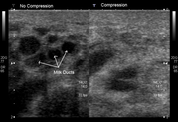
Figure 3.

Cross-sectional ultrasound image of milk ducts in the lactating breast. On the left image, milk ducts appear as oval hypoechoic (black) structures. On the right image, milk ducts have collapsed under minimal to moderate compression with the ultrasound transducer.
