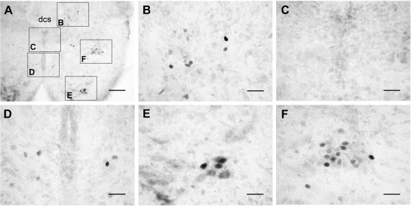
Figure 4.
In vivo c-Fos expression in the cord. Basal levels of c-Fos expression under normal physiological conditions were promptly preserved by a transcardial perfusion of fixative. (A) Photomicrograph of a transverse section of the T3 spinal cord segment. Insets in (A) are magnified as indicated. c-Fos-positive nuclei were distributed in the dorsal laminae near the dcs (B), around the cc (D), in the ventral horn (E), and in the IML (F). No c-Fos-positive cells were found in lamina X (C), the lfu, or the outer rim of the white matter (A). Scale bars = 100 μm in (A) and 25 μm in (B-F).

