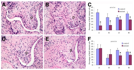
Fig. 5.
Osr2-IresCre;Smoc/c mutant embryos exhibited defects in palatal shelf growth at E13.5. Frontal sections through the palatal regions of BrdU-labeled Osr2-IresCre;Smo+/c (A,D) and Osr2-IresCre;Smoc/c mutant (B,E) embryos were stained with anti-BrdU antibody. The labeled cell nuclei were stained blue. The white line in each panel divided each image of the palatal shelf to medial and lateral halves for the calculation of the percentage of BrdU-labeled nuclei in those regions separately. A and B show typical sections through the middle of the anterior half of the palatal shelves, whereas D and E are from the posterior third of the palatal shelves. (C,F) Comparison of the percentage of BrdU-labeled cells in the anterior (C) and posterior (F) regions of the developing palate in the Osr2-IresCre;Smo+/c (control) and Osr2-IresCre;Smoc/c (mutant) embryos. Standard deviation values were used for the error bars. An asterisk denotes a significant reduction in the percentage of BrdU-labeled cells in the mutant (P<0.05). p, palatal shelf.

