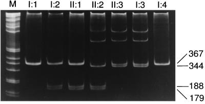
Figure 5.
Confirmation and segregation of HYAL1 mutations. Fragment C (Fig. 2) was PCR-amplified from genomic DNA, was digested with MscI, and was separated by PAGE. The 1412G → A mutation creates an MscI site that generates 188- and 179-bp fragments from the 367-bp PCR product, as seen in the patient (II:2), her father (II:1), and her paternal grandmother (I:2). The two slower-migrating bands in lanes II:2, II:3, and I:3 are heteroduplexes formed between the normal (367 bp) and 1361del37ins14 (344 bp) PCR products. The sizes of the bands are shown to the right of the gel. The numbering refers to the pedigree in Fig. 1.

