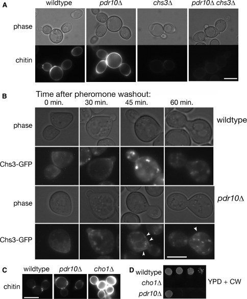
Fig. 4.
Defective endocytosis of Chs3 causes excess chitin deposition in pdr10∆ cells. a Cells from log phase cultures of strains W303-1A (wild type), NRY243 (pdr10∆), NRY602 (chs3∆), and NRY601 (pdr10∆ chs3∆) were stained for chitin and examined by fluorescence microscopy. Bar, 4 μm. b Log phase cells from strains W303-1A and NRY251 (pdr10∆) carrying plasmids pRS315-CHS7 and pCRP12 were treated with 3.6 μM α-factor for 35 min in SCD buffered to pH 3.5 with 25 mM sodium succinate and supplemented with adenine. Pheromone was then washed out at time 0, and the localization of Chs3-GFP was monitored by fluorescence microscopy as described under Materials and Methods. Punctate Chs3-GFP staining of the plasma membrane in pdr10∆ cells at late time points is indicated by arrow heads. Bar, 3 μm. c Strains W303-1A, NRY243, and NRY407 (cho1∆) were grown into log phase, stained for chitin, and examined by fluorescence microscopy. Bar, 5 μm. d Serial dilutions of overnight cultures of strains W303-1A, NRY243, and NRY407 were spotted onto YPD plates containing Calcofluor

