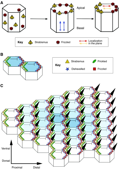
Fig. 2.
PCP establishment in the Drosophila wing. (A) Polarity establishment in the Drosophila wing epithelium as a classic model of PCP. PCP establishment first requires key proteins [e.g. Strabismus (Stbm) and Frizzled (Fz)] to be apically distributed and then localized to the proximal and distal sides of the apical surface, respectively, in the plane that lies orthogonal to the apical-basal axis. See Zallen (Zallen, 2007). (B) Stbm then recruits Prickled to the proximal side of the cell, which antagonizes Dishevelled (Dsh), hence restricting the location of the Dsh-Fz complex to the opposite, distal side. At the same time, interactions between the extracellular domains of Stbm and Fz at adjacent proximal and distal membranes of neighboring cells stabilize the polarized configurations, allowing a small clone of cells to be polarized. (C) A speculative model of PCP (see Tree et al., 2002b), in which the organized asymmetric distribution of core PCP proteins spreads from a small group of polarized organizer cells. A wave of polarized interactions emanates from an initial site of polarization outwards in all directions through the plane of the tissue, establishing a coordinated array of polarized cells (indicated here by the decreasing intensity of blue shading from the center to outer cells). See Jones and Chen (Jones and Chen, 2007).

