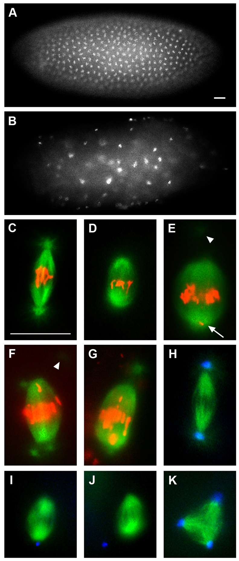
Fig. 1.

The nopo phenotype. Representative syncytial embryos and mitotic spindles in embryos from wild-type or nopoZ1447 females. (A,B) Staining of nopo-derived embryos reveals developmental arrest with condensed, unevenly spaced DNA (B) compared with wild type (A). (C-G) Microtubules are in green and DNA in red. (C) Wild-type spindle. (D-F) Shortened, barrel-shaped nopo spindles with detached centrosomes and misaligned chromosomes. Arrowheads indicate detached centrosomes out of focal plane; arrow, DNA at pole. Metaphase-like spindle with two centrosomes per pole (F) reveals an asynchrony of nuclear and centrosome cycles. (G) Similar defects are observed in an nopoExc142/Df(2R)Exel7153-derived embryo. (H-K) Microtubules are in green; centrosomes in blue. (H) Wild-type spindle. (I,J) nopo spindles with detached and/or missing centrosomes. (K) Tripolar nopo spindle. Scale bars: 20 μm.
