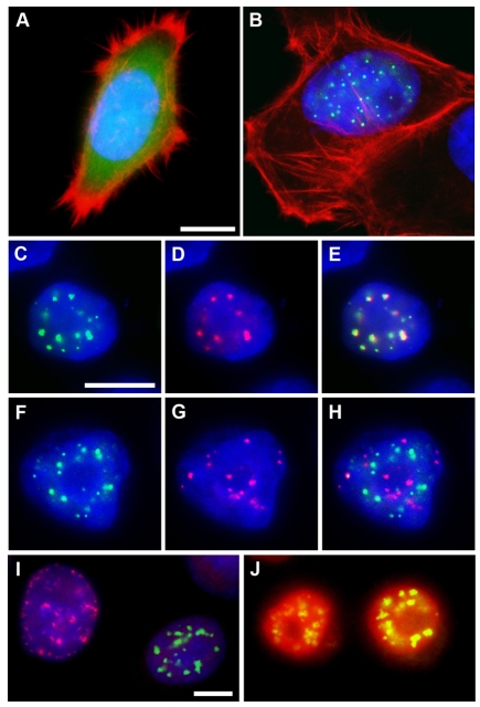
Fig. 5.
Nuclear localization of NOPO. Immunofluorescence microscopy of transfected HeLa cells. DNA is in blue. (A,B) eGFP is in green and actin in red. eGFP-Drosophila NOPO (B) localizes to nuclear puncta; eGFP (A) is homogeneously distributed. (C-E) eGFP is in green and mCherry in red. eGFP-Drosophila NOPO (C) and mCherry-human TRIP (D) co-localize in nuclear puncta (E, merge). (F-H) eGFP is in green and CREST in red. eGFP-NOPO (F) is not at the centromeres (G; H, merge). (I) Cells with eGFP-NOPO puncta (green) are negative for PCNA puncta (red). (J) Cells with eGFP-NOPO puncta (green) are positive for nuclear Cyclin A (red). Scale bars: 10 μm.

